
Cartilage: Types, Structure & Functions | MBBS Anatomy
Cartilage is a specialized connective tissue that provides support, flexibility and smooth surfaces for movement. It is an important component of the developing skeleton and remains present in several structures of the adult body.
There are three major types of cartilage: hyaline cartilage, elastic cartilage and fibrocartilage. They differ in their matrix, fiber composition, mechanical properties and distribution.
Interactive Cartilage Guide
Click the buttons below to compare the major types of cartilage and view their microscopic appearance.
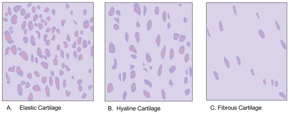
What Is Cartilage?
Cartilage is a specialized form of connective tissue composed of chondrocytes embedded within an extracellular matrix.
The matrix contains collagen fibers, proteoglycans, water and other components that determine the mechanical properties of the tissue.
Unlike bone, cartilage does not contain blood vessels within its matrix. Nutrients reach chondrocytes mainly by diffusion through the matrix.
Cells of Cartilage
Chondroblasts
Chondroblasts are developing cartilage-forming cells. They produce components of the cartilage extracellular matrix.
Chondrocytes
Chondrocytes are mature cartilage cells. They are located within spaces called lacunae and maintain the surrounding matrix.
Basic Structure of Cartilage
Cartilage consists mainly of cells and extracellular matrix.
- Chondrocytes are located in lacunae.
- The extracellular matrix contains water, proteoglycans and collagen fibers.
- The type and arrangement of fibers vary between the three types of cartilage.
- Cartilage is usually surrounded by a connective tissue layer called the perichondrium.
Types of Cartilage
| Type | Main Feature | Important Examples |
|---|---|---|
| Hyaline cartilage | Smooth matrix with mainly type II collagen. | Articular surfaces, costal cartilages, respiratory tract, nasal cartilage and epiphyseal plates. |
| Elastic cartilage | Contains type II collagen and abundant elastic fibers. | External ear, epiglottis and parts of the larynx. |
| Fibrocartilage | Contains abundant type I collagen and is highly resistant to tensile and compressive forces. | Intervertebral discs, pubic symphysis and menisci. |
Hyaline Cartilage
Hyaline cartilage is the most widely distributed type of cartilage in the body.
Its matrix has a relatively smooth and glass-like appearance because the collagen fibers are fine and not easily visible by routine microscopy. The principal collagen is type II collagen.
Functions of Hyaline Cartilage
- Provides smooth articular surfaces at synovial joints.
- Provides structural support to the respiratory tract.
- Forms the costal cartilages connecting ribs to the sternum.
- Provides a temporary skeletal model during endochondral ossification.
- Forms the epiphyseal growth plate during skeletal growth.
Locations of Hyaline Cartilage
- Articular surfaces of synovial joints
- Costal cartilages
- Nose
- Trachea
- Bronchi
- Parts of the larynx
- Epiphyseal plates
Elastic Cartilage
Elastic cartilage resembles hyaline cartilage but contains a prominent network of elastic fibers within its matrix.
The elastic fibers allow the tissue to maintain its shape while providing greater flexibility than ordinary hyaline cartilage.
Functions
- Provides flexible support.
- Maintains the shape of certain structures.
- Allows bending and recoil.
Locations
- External ear
- Epiglottis
- Auditory tube
- Some cartilages of the larynx
Fibrocartilage
Fibrocartilage is the strongest type of cartilage in terms of resistance to combined tensile and compressive forces.
It contains abundant bundles of type I collagen. The chondrocytes are commonly arranged between the collagen bundles.
Functions
- Resists compression.
- Resists tensile forces.
- Provides strong structural support.
- Helps absorb and distribute mechanical loads.
Locations
- Intervertebral discs
- Pubic symphysis
- Menisci of the knee
- Articular discs in certain joints
Hyaline vs Elastic vs Fibrocartilage
| Feature | Hyaline | Elastic | Fibrocartilage |
|---|---|---|---|
| Main collagen | Type II | Type II | Type I, with type II also present |
| Elastic fibers | Absent as a prominent component | Abundant | Not a characteristic component |
| Matrix | Smooth and glassy | Flexible matrix with elastic fibers | Dense collagen-rich matrix |
| Strength | Moderate support | Flexible support | High resistance to tensile and compressive forces |
| Perichondrium | Usually present | Present | Absent |
| Major examples | Articular cartilage, trachea, costal cartilage | External ear, epiglottis | Intervertebral discs, menisci, pubic symphysis |
Perichondrium
The perichondrium is a layer of connective tissue that surrounds most cartilage.
It contains blood vessels that supply nutrients to the underlying cartilage by diffusion.
The perichondrium has an outer fibrous layer and an inner chondrogenic layer containing cells capable of contributing to cartilage growth.
- Articular cartilage does not have a perichondrium.
- Fibrocartilage does not have a perichondrium.
Growth of Cartilage
Cartilage can grow by two mechanisms: interstitial growth and appositional growth.
Interstitial Growth
Interstitial growth occurs from within the cartilage. Chondrocytes divide and produce additional matrix, causing the cartilage to expand internally.
Appositional Growth
Appositional growth occurs by addition of new cartilage to the surface. Cells in the inner layer of the perichondrium can differentiate into chondroblasts and contribute to new cartilage formation.
Cartilage and Bone: Important Difference
| Feature | Cartilage | Bone |
|---|---|---|
| Blood supply | Avascular | Vascular |
| Main cells | Chondrocytes | Osteocytes |
| Matrix | Flexible, hydrated extracellular matrix | Mineralized extracellular matrix |
| Repair | Generally limited | Usually better than cartilage |
| Main role | Support, flexibility and smooth surfaces | Support, protection, movement and mineral storage |
Clinical Importance
Articular Cartilage Damage
Articular cartilage has a limited capacity for repair. Damage to the cartilage covering a joint surface can therefore produce persistent structural abnormalities and contribute to degenerative joint disease.
Osteoarthritis
Degeneration of articular cartilage is an important feature of osteoarthritis. Loss of the smooth cartilage surface can increase friction and alter joint mechanics.
Intervertebral Disc
The intervertebral disc contains fibrocartilage within the annulus fibrosus and plays an important role in distributing mechanical loads between adjacent vertebrae.
Growth Plate
The epiphyseal plate is composed of hyaline cartilage and is responsible for longitudinal growth of a growing long bone.
Cartilage Repair
Because cartilage is avascular, repair is generally slower and less complete than repair in many vascular tissues.
High-Yield MBBS Points
- There are three major types of cartilage: hyaline, elastic and fibrocartilage.
- Hyaline cartilage mainly contains type II collagen.
- Elastic cartilage contains type II collagen plus elastic fibers.
- Fibrocartilage contains abundant type I collagen.
- Cartilage is generally avascular and aneural.
- Chondrocytes are located in lacunae.
- The perichondrium surrounds most cartilage.
- Articular cartilage lacks a perichondrium.
- Fibrocartilage lacks a perichondrium.
- Hyaline cartilage forms the epiphyseal growth plate.
- Fibrocartilage is found in intervertebral discs and menisci.
- Elastic cartilage is found in the external ear and epiglottis.
Quick Revision Table
| Cartilage | Key Word | Examples |
|---|---|---|
| Hyaline | Type II collagen | Articular surface, trachea, costal cartilage |
| Elastic | Elastic fibers | External ear, epiglottis |
| Fibrocartilage | Type I collagen | Intervertebral discs, menisci, pubic symphysis |
Summary
Cartilage is a specialized connective tissue composed of chondrocytes embedded in an extracellular matrix. It provides support, flexibility, shock absorption and smooth surfaces for movement.
The three major types are hyaline cartilage, elastic cartilage and fibrocartilage. Hyaline cartilage is widely distributed and is important in joints, respiratory structures, costal cartilages and growth plates.
Elastic cartilage provides flexible support, while fibrocartilage provides strong resistance to tensile and compressive forces. Cartilage is generally avascular and therefore has limited capacity for repair.
Image Credits & Licenses
1. Three Types of Cartilage:
Shiloh117981894, Cartilage types.jpg.
Licensed under CC BY-SA 4.0.
Wikimedia Commons
2. Hyaline Cartilage:
Berkshire Community College Bioscience Image Library,
Connective Tissue Hyaline Cartilage (26989335717).jpg.
Licensed under CC0 1.0.
Wikimedia Commons
3. Elastic Cartilage:
Echinaceapallida, Cartilage elastic1.jpg.
Licensed under CC BY-SA 4.0.
Wikimedia Commons
4. Fibrocartilage:
Berkshire Community College Bioscience Image Library,
Connective Tissue Fibrocartilage (27988651538).jpg.
Licensed under CC0 1.0.
Wikimedia Commons
Medical Disclaimer: The information provided in this article is strictly for educational, study, and exam-preparation purposes. It does not constitute professional medical advice, diagnosis, or treatment. Always consult a qualified healthcare provider for clinical decisions.