
Bone vs Cartilage: Structure, Functions & Differences | MBBS Anatomy
Bone and cartilage are specialized connective tissues that form important parts of the human skeletal system. Although both provide support and contribute to movement, they differ significantly in their matrix, blood supply, cells, mechanical properties and ability to repair.
Understanding these differences is important for MBBS anatomy, histology, orthopedics and clinical medicine.
Interactive Bone vs Cartilage Guide
Use the buttons below to view the main microscopic features and compare bone with cartilage.
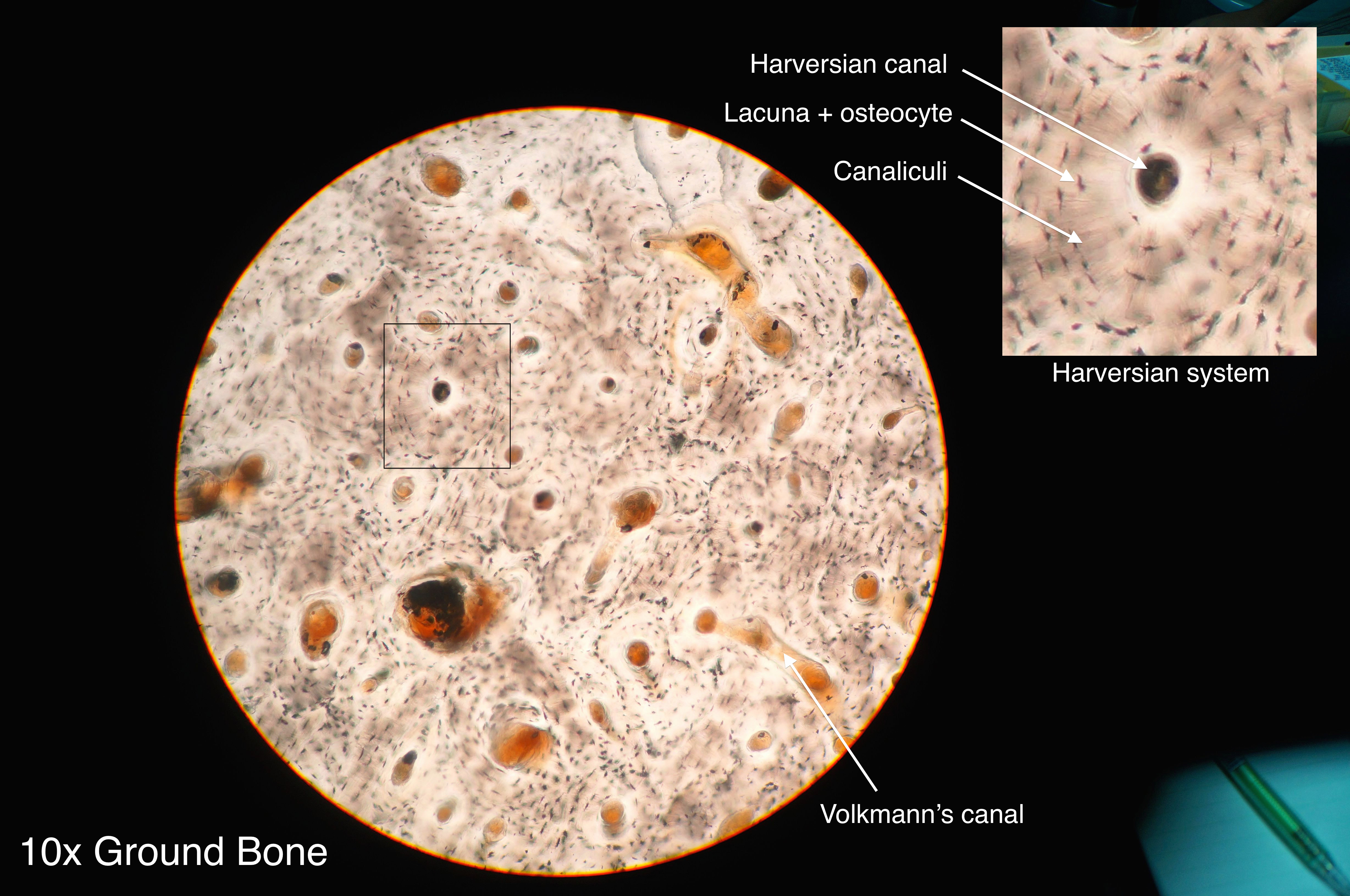
What Is Bone?
Bone is a specialized mineralized connective tissue that forms the rigid framework of the skeleton.
It provides mechanical support, protects internal organs, provides attachment sites for muscles and tendons, participates in movement and serves as a major reservoir for minerals such as calcium and phosphate.
Main Cells of Bone
- Osteoblasts: bone-forming cells that synthesize osteoid.
- Osteocytes: mature bone cells located in lacunae.
- Osteoclasts: multinucleated cells responsible for bone resorption.
What Is Cartilage?
Cartilage is a specialized connective tissue in which chondrocytes are embedded within an abundant extracellular matrix.
It provides flexible support, smooth articulating surfaces and resistance to compression. Cartilage is generally avascular, so nutrients reach its cells mainly by diffusion through the matrix.
Main Cells of Cartilage
- Chondroblasts: immature cartilage-forming cells.
- Chondrocytes: mature cartilage cells located in lacunae.
Bone vs Cartilage: Major Differences
| Feature | Bone | Cartilage |
|---|---|---|
| Nature of tissue | Hard, mineralized connective tissue | Firm but flexible connective tissue |
| Main cells | Osteoblasts, osteocytes and osteoclasts | Chondroblasts and chondrocytes |
| Cells in lacunae | Osteocytes | Chondrocytes |
| Matrix | Mineralized extracellular matrix | Hydrated, flexible extracellular matrix |
| Major collagen | Type I collagen | Usually type II collagen; fibrocartilage has abundant type I collagen |
| Blood supply | Vascular | Generally avascular |
| Nerve supply | Bone and periosteum contain nerve fibers | Cartilage itself is generally aneural |
| Perichondrium | Not applicable | Present around most cartilage; absent from articular cartilage and fibrocartilage |
| Mechanical property | Rigid and strong | Firm, flexible and compression-resistant |
| Repair | Generally better repair capacity | Generally limited repair capacity |
Structure of Bone
Bone consists of cells embedded within a mineralized extracellular matrix. Its organization allows it to withstand substantial mechanical loads.
Compact Bone
Compact bone forms the dense outer component of most bones. Its microscopic structural unit is the osteon, also called the Haversian system.
An osteon contains concentric lamellae surrounding a central Haversian canal, which carries blood vessels and nerves. Osteocytes occupy lacunae and communicate through tiny canaliculi.
Cancellous Bone
Cancellous bone, also called trabecular or spongy bone, consists of interconnected trabeculae with spaces between them. These spaces may contain bone marrow.
Structure of Cartilage
Cartilage has chondrocytes embedded within an extracellular matrix. The composition of this matrix determines the mechanical properties of each type of cartilage.
Hyaline Cartilage
Hyaline cartilage contains mainly type II collagen and has a relatively smooth, glass-like matrix.
Elastic Cartilage
Elastic cartilage contains type II collagen together with abundant elastic fibers. It provides flexible support.
Fibrocartilage
Fibrocartilage contains abundant type I collagen arranged in bundles and is particularly adapted to resist tensile and compressive forces.
Why Is Bone Harder Than Cartilage?
The major reason is the difference in their extracellular matrices. Bone matrix becomes mineralized with calcium-phosphate-containing mineral, which gives bone its characteristic rigidity.
Cartilage has a highly hydrated matrix containing proteoglycans and collagen fibers. This gives it a combination of firmness, flexibility and resistance to compression.
Blood Supply
Bone
Bone is vascular. Blood reaches bone through nutrient arteries, periosteal vessels and vessels supplying metaphyseal and epiphyseal regions. Compact bone also contains vascular channels within its microscopic architecture.
Cartilage
Cartilage itself is generally avascular. When a perichondrium is present, blood vessels within the perichondrium provide a source of nutrients that diffuse through the cartilage matrix.
Articular cartilage is a special case because it lacks a perichondrium and is nourished largely by diffusion from synovial fluid and adjacent tissues.
Growth of Bone vs Cartilage
| Feature | Bone | Cartilage |
|---|---|---|
| Growth in length | Long bones grow longitudinally at the epiphyseal plate. | Cartilage can grow by interstitial growth. |
| Growth in thickness | Occurs through appositional growth and remodeling. | Can occur by appositional growth from the perichondrium. |
| Remodeling | Continuous throughout life. | Much more limited than bone remodeling. |
Bone and Cartilage in the Developing Skeleton
Cartilage plays an important role during skeletal development. In endochondral ossification, a hyaline cartilage model is progressively replaced by bone.
This mechanism is responsible for formation of most long bones and contributes to longitudinal skeletal growth through the epiphyseal plate.
Cartilage is not simply "converted into bone." During endochondral ossification, a cartilage model is progressively remodeled and replaced by bone tissue.
Clinical Importance
Fracture of Bone
Bone has a relatively good capacity for repair because it is vascular and contains active bone-forming and remodeling cells.
Articular Cartilage Damage
Articular cartilage has limited intrinsic repair capacity because it is avascular and lacks a perichondrium. Damage can therefore persist and may contribute to degenerative joint disease.
Osteoarthritis
Osteoarthritis involves structural changes in synovial joints, including damage and loss of articular cartilage along with changes in underlying bone and other joint tissues.
Intervertebral Disc
The annulus fibrosus of the intervertebral disc contains fibrocartilage and helps the disc resist mechanical forces.
Growth Plate Disorders
Because the epiphyseal plate is made of hyaline cartilage, disorders affecting cartilage growth or mineralization can interfere with longitudinal bone growth.
Bone vs Cartilage: Quick Comparison
| Bone | Cartilage |
|---|---|
| Hard and rigid | Firm and flexible |
| Mineralized matrix | Hydrated extracellular matrix |
| Vascular | Generally avascular |
| Osteocytes in lacunae | Chondrocytes in lacunae |
| Type I collagen predominates | Type II collagen predominates in most cartilage |
| Forms the main adult skeleton | Provides flexible support and specialized joint surfaces |
| Relatively good repair capacity | Limited repair capacity |
High-Yield MBBS Points
- Bone is a mineralized connective tissue.
- Cartilage is a specialized connective tissue with an abundant extracellular matrix.
- Osteocytes are the mature cells of bone.
- Chondrocytes are the mature cells of cartilage.
- Both osteocytes and chondrocytes are found in lacunae.
- Bone is vascular.
- Cartilage is generally avascular and aneural.
- Type I collagen predominates in bone matrix.
- Type II collagen predominates in hyaline and elastic cartilage.
- Fibrocartilage contains abundant type I collagen.
- Articular cartilage lacks a perichondrium.
- Fibrocartilage also lacks a perichondrium.
- Bone undergoes continuous remodeling throughout life.
- Cartilage generally has a more limited capacity for repair.
One-Minute Revision
| Question | Answer |
|---|---|
| Which tissue is mineralized? | Bone |
| Which tissue is generally avascular? | Cartilage |
| Mature cell of bone? | Osteocyte |
| Mature cell of cartilage? | Chondrocyte |
| Cells of bone lie in? | Lacunae |
| Cells of cartilage lie in? | Lacunae |
| Main collagen of bone? | Type I |
| Main collagen of hyaline cartilage? | Type II |
| Cartilage at synovial articular surfaces? | Hyaline cartilage |
| Cartilage in intervertebral discs? | Fibrocartilage |
Summary
Bone and cartilage are both specialized connective tissues, but their structure and mechanical properties are different.
Bone has a mineralized matrix, is vascular and provides a rigid framework for support, protection and movement.
Cartilage has a flexible extracellular matrix, is generally avascular and provides support, flexibility, smooth joint surfaces and resistance to compression.
For MBBS examinations, remember the key distinction: bone is vascular and mineralized, whereas cartilage is generally avascular and has a flexible matrix.
Image Credits & Licenses
1. Compact Bone Histology:
Athikhun.suw, Compact bone histology 2014.jpg.
Licensed under CC BY-SA 4.0.
Wikimedia Commons
2. Hyaline Cartilage Histology:
Berkshire Community College Bioscience Image Library,
Connective Tissue Hyaline Cartilage (26989335717).jpg.
Licensed under CC0 1.0 / Public Domain Dedication.
Wikimedia Commons
Medical Disclaimer: The information provided in this article is strictly for educational, study, and exam-preparation purposes. It does not constitute professional medical advice, diagnosis, or treatment. Always consult a qualified healthcare provider for clinical decisions.